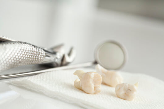
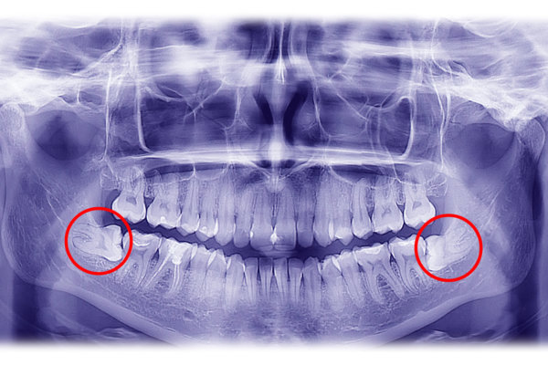
Wisdom teeth, or third molars are one of the most frequent causes of dental pain. The lack of space or malposition of them can cause that they get impacted (or 'trapped') within the jaws, causing:
- caries in contiguous teeth
- inflammation of the gums
- maxillary cists
Antibiotics and analgesics can soothe the acute pain they can cause, but if we do not extract them, the problem is still present.
Wisdom teeth are surrounded by anatomical structures, and it is very important to extract them without damaging them. Near the upper wisdom teeth we find the maxillary sinuses and in the vicinity of the lower ones we find the dental nerve (which gives us sensitivity to the teeth and lower lip) and the lingual nerve (it provides sensitivity to the tongue). It is, therefore, of vital importance to put yourself in experienced hands, to minimize the risks of affecting these nerves.

For proper diagnosis and planning of wisdom teeth extraction, it is important to have adequate radiological images that accurately report the size , form, and situation in relation to the rest of the structures. At the Maxillofacial Institute we generated these images with an I-CAT device. It is basically a three-dimensional low-radiation scanner that provides all the necessary information to minimize the risks of extraction, identifying structures adjacent to the tooth, and allowing proper planning.
Wisdom tooth extraction procedure is carried out for the most part of the cases with local anesthesia. For cases in which it is necessary to extract more than one tooth, or that they are in a difficult position, we use intravenous sedation techniques administered by our anesthetist. These allow to eliminate the anxiety component of the patient and make the surgical experience completely painless and anxiety-free.
It might interest you: Should I get my wisdom teeth removed even if they do not bother me?
Related: What to eat after your wisdom tooth extraction?


